


WHO 2008 diagnosis standard of tumors of hematopoietic and lymphoid tissues: according to morphology(M), immunology (I), cytogenetics(C) and molecular biology (M) comprehensive diagnosis.
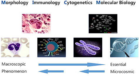
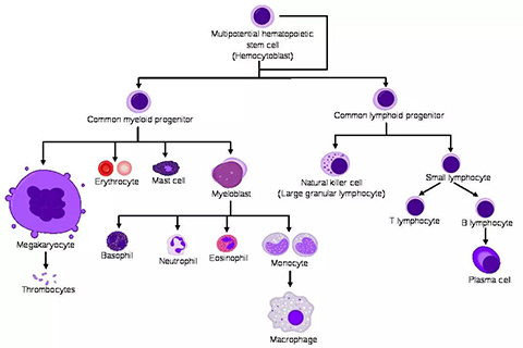
Blood disease often have abnormal blood cell morphology, to accurately identify the abnormal cell morphology is very need to experience things.
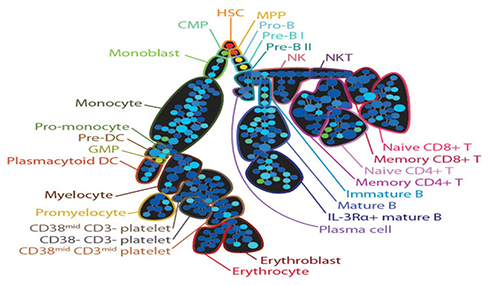
Flow cytometry mainly uses antigen binding antibody and fluorescence labeling principle to make quantitative analysis of multiple parameters of cells, so it is often called immuno-typing. But flow can do more than that. Flow analysis is highly dependent on knowledge and experience.
There are nearly 30,000 specimens per year, and 50% of them are from all over China. This lab has become a well-known center for diagnosis of difficult diseases.
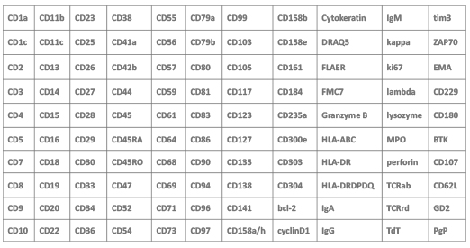
There are over 110 antibodies of different coding in the lab.
Cytogenetic/Fluorescence in Situ Hybridization (FISH) Lab
More than 10,000 specimens were received every year, of which more than 1/3 were specimens from other hospitals, including many complicated karyotypes and difficult cases. Our time of chromosome presentation is only 6-8 working days.
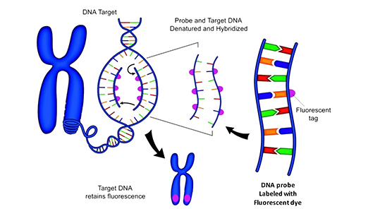
There are some subtle chromosomal abnormalities, and karyotype analysis is difficult to distinguish, and it needs to be detected by FISH method.
In our lab, there are more than 400 FISH test species.

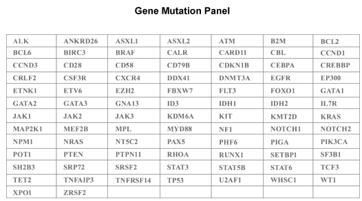
Most tumors are the result of a combination of multiple gene mutations. It's important to understand it in order to treat the tumor.
Gene Mutation Panel: includes hot-spot mutation zone or whole-length coding region of 86 genes.
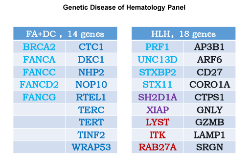
Genetic factors of some genetic diseases may have complicated relationships with a variety of clinical syndromes.
Genetic Disease of Hematoloy Panel: about Fanconi anemia (FA) & Congenital dyskeratosis (DC) are 14 genes, about Hemophagocyic histiocytosis (HLH) are 18 genes.
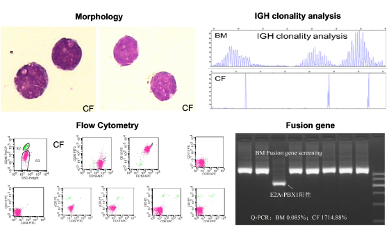
Cerebrospinal fluid specimens from a patient with central nervous system symptoms (who had been diagnosed as tuberculous meningitis for 4+ months) were successfully examined by MICM.
Patient No.WJJ, female, 21 years old, onset with central nervous system symptoms, without lymphadenopathy or hepatosplenomegaly. She had sought for diagnosis and treatment at neurology department in several hospitals.
Cerebrospinal fluid (CF) sample was sent to our laboratory to do morphology and flow cytometry testing at 14.Aug.2008. Cytogenetic and genetic testing were added based on the morphology and immunophenotyping results. Bone marrow (BM) biopsy was then acquisited in the same day and inspected.
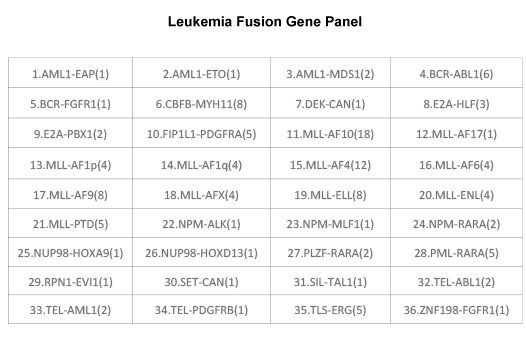
According to the examination results, it was diagnosed as "primary lymphoblastic lymphoma with t (1,19) (q23,P13) translocation and E2A-PBX1 fusion gene". This is not a nervous system disease.
Cerebrospinal fluid specimens from a patient with central nervous system symptoms (who had been diagnosed as tuberculous meningitis for 4+ months) were successfully examined by MICM.
Cerebrospinal fluid specimens from a patient with central nervous system symptoms (who had been diagnosed as tuberculous meningitis for 4+ months) were successfully examined by MICM.
Fatient No.WJJ, female, 21 years old, onset with central nervous system symptoms, without lymphadenopathy or hepatosplenomegaly. She had sought for diagnosis and treatment at neurology department in several hospitals.
Cerebrospinal fluid (CF) sample was sent to our laboratory to do morphology and flow cytometry testing at 14.Aug.2008. Cytogenetic and genetic testing were added based on the morphology and immunophenotyping results. Bone marrow (BM) biopsy was then acquisited in the same day and inspected.
According to the examination results, it was diagnosed as "primary lymphoblastic lymphoma with t (1,19) (q23,P13) translocation and E2A-PBX1 fusion gene". This is not a nervous system disease.
The patient received intrathecal injection with MTX+IDA and Dex+L-ASP chemotherapy, from the day after chemotherapy her headache disappeared, and her lower extremity muscle strength recovered to normal on the 15 days after chemotherapy.
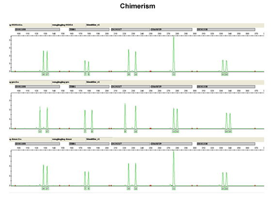
After one course of chemotherapy, allo-HSCT treatment with radiotherapy was continued.
The chimerism rate of patients reached the full donor origin which checked by 1 month after HSCT. It means her hematopoietic system has been rebuilt.
Until now, she achieved continuous remission.


 京公网安备13108202000843号